

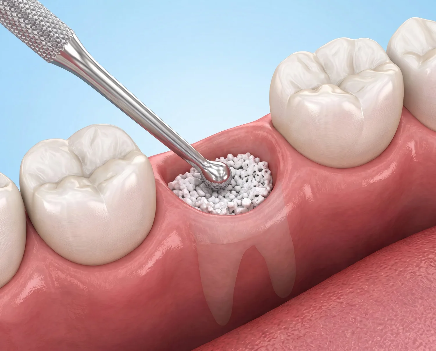
What Is an Autogenous Tooth Graft?
An autogenous tooth graft is graft material obtained from a person’s own extracted tooth. Dentin, the hard tissue of the tooth, is structurally similar to bone: both consist largely of collagen and calcium phosphate compounds. This similarity allows properly processed dentin to act as a scaffold that can replace bone.
The extracted tooth is converted into particles in a specialized grinding device during the same session, then soaked in a cleaning solution to remove bacteria and rinsed. The resulting material is applied to the same patient during the same session. Because the tissue comes from the patient, the immune system does not recognize it as foreign.
How Can an Extracted Tooth Be Used?
1. Socket preservation: After a tooth is extracted, the surrounding bone gradually begins to resorb. When processed dentin is placed in the socket, it helps preserve bone volume. This is important in areas where an implant may be planned in the future.
2. Increasing bone volume: In areas with insufficient bone, autogenous dentin graft can be used as bone graft material. It may also be used in procedures such as sinus floor elevation.
3. Repairing gum and bone defects: It may be used to fill defects caused by gum disease or infection-related bone loss.
4. Tooth autotransplantation: In some cases, the extracted tooth itself can be transplanted into another space in the mouth. The most common example is placing an unused wisdom tooth in the position of a lost molar.
5. Storage of dental pulp tissue: Because the pulp tissue inside the tooth contains stem cells, it can be frozen and stored at authorized tissue banks. This is not a treatment, but a storage procedure. The conditions in which it may be used are still under investigation.
Which of these options is appropriate depends on the condition of the tooth. Teeth with extensive decay, fractures, or infection may not be suitable for every method.
Who Is It Suitable For?
Evaluation may be considered in the following situations: an implant is planned in the area of the tooth to be extracted, bone support may be needed in another area when a wisdom tooth is extracted, a bone defect is present due to gum disease, a healthy tooth needs to be extracted for orthodontic reasons, or a suitable tooth is available for transplantation in place of a lost molar.
There are also situations in which these methods are not suitable. An active infection in the mouth, fragmentation of the tooth during extraction, extensive destruction of the dentin, or a systemic condition that affects bone healing may limit their use. The decision is made after a clinical examination and imaging.
Treatment Process
1. Planning: The tooth to be extracted and the treatment area are evaluated with X-rays and, when necessary, three-dimensional tomography. The appropriate method is decided at this stage.
2. Extraction: The tooth is extracted while preserving the surrounding bone. Fillings, crowns, and soft tissue remnants are removed from the tooth.
3. Processing: The tooth is converted into particles in a grinding device, soaked in a cleaning solution, and rinsed. This stage takes approximately fifteen minutes.
4. Application: The prepared material is placed in the planned area, covered with a membrane when necessary, and the area is sutured.
5. Healing: Swelling and mild pain are expected findings during the first few days. Bone maturation progresses over several months. In areas where an implant is planned, this period is monitored through follow-up examinations.
Who Carries Out Your Treatment?
At Papatya Dental Clinic, this treatment is carried out by specialists in oral and maxillofacial surgery and periodontology:
- Ozan Ergen, Specialist in Oral, Dental and Maxillofacial Surgery: Hacettepe University, specialist training at Dicle University, 16 years of experience, member of ITI (International Team for Implantology)
- Ali Bulut, Specialist Oral, Dental and Maxillofacial Surgeon: Ankara University, specialist training at Süleyman Demirel University, 10 years of experience, member of ITI
- Assoc. Prof. Gülen Kamak, Specialist in Periodontology and Implantology: Selçuk University, doctoral and specialist training at Atatürk University, 20 years of experience, member of ITI and the European Federation of Periodontology
- Elif Teke, Specialist Periodontist: Ankara University, specialty training at Süleyman Demirel University, 13 years of experience, member of ITI
Planning, laboratory and implant systems
- 3D CT (CBCT) scan to measure bone height and width before surgery
- Grafting and the later implant surgery are carried out by the same surgical team
- A wide choice of implant systems, including Straumann, Nobel Biocare, Neodent, Osstem, MegaGen and Dentium for the implant stage
- You receive an implant card for every implant placed. The warranty period depends on the implant system used.
Which treatment suits you is decided after a clinical examination and a CT scan.