Papatya Dental7/24

About this page

This page explains what we use each device for. Which device is needed for a particular procedure is decided by the dentist after your examination.

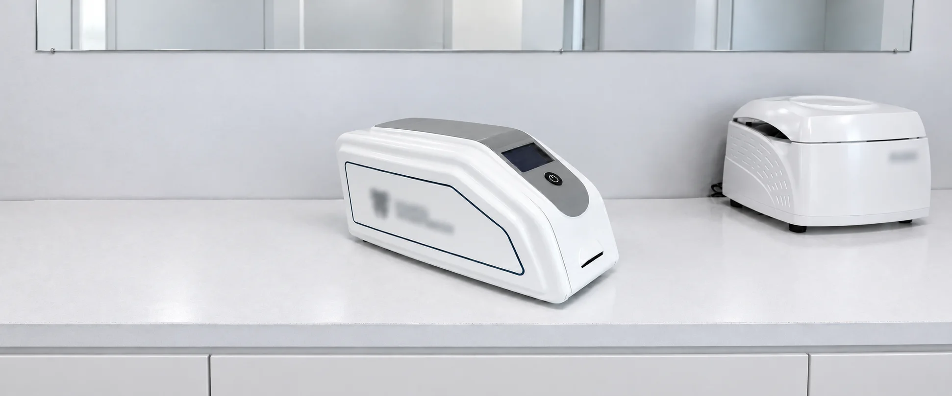
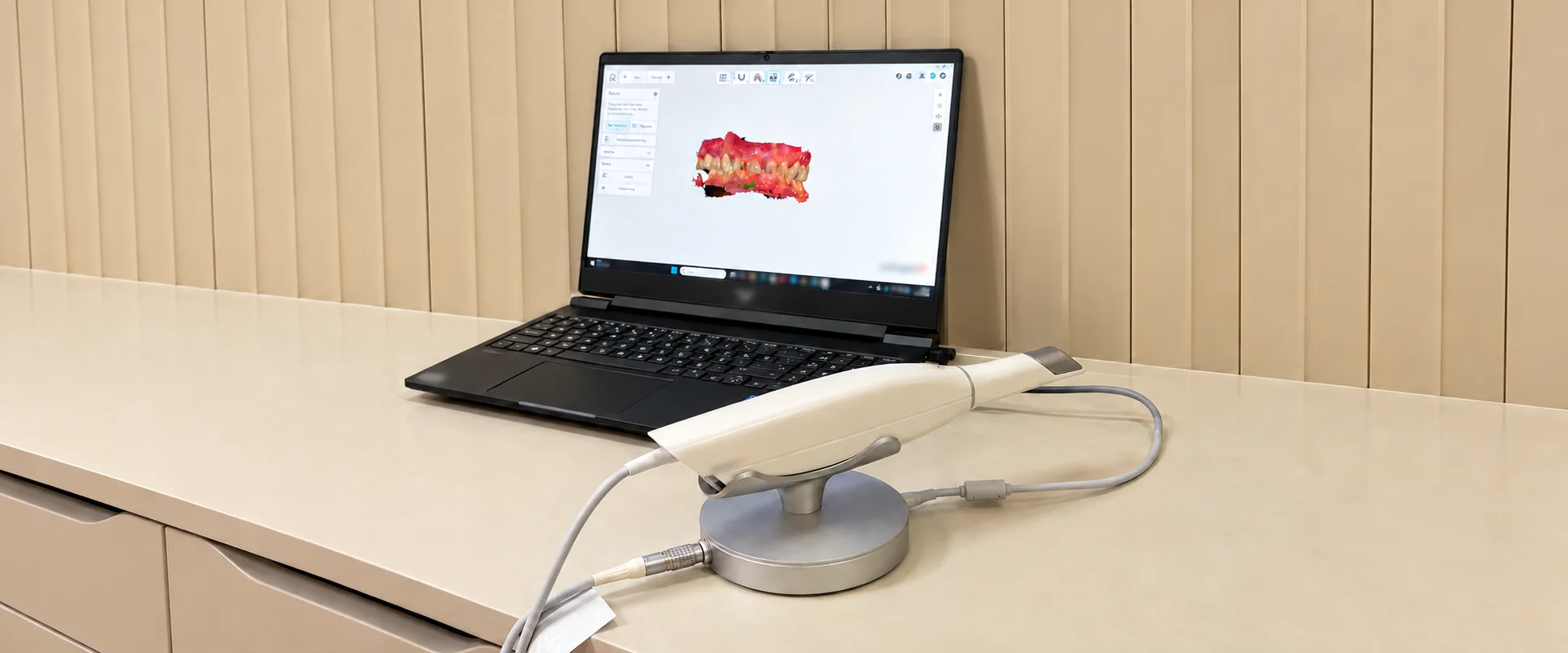
Intraoral Digital Scanner

How are digital impressions of oral surfaces taken?

Optical data from teeth and nearby surfaces form a 3D model. In suitable cases, this record can support restoration planning and laboratory work. The dentist selects the impression method; scanning does not replace all other impression and imaging methods.

  • Recording teeth and nearby surfaces
  • Three-dimensional digital model
  • Digital data sharing with the laboratory

Intraoral Digital Scanner
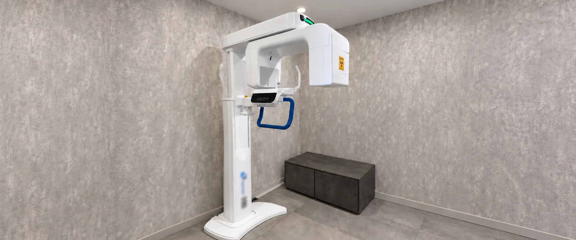
When is three-dimensional imaging of the jaws and teeth needed?

When examination and existing images are insufficient, 3D cross-sectional imaging may be requested if needed. The dentist assesses bone and nearby anatomy together. It is not routine for every patient; as it uses X-rays, the decision is made individually.

  • Bone height
  • Bone width
  • Nerve canal position
  • Impacted tooth position

Cone Beam Computed Tomography

CBCT

Cone Beam Computed Tomography — CBCT

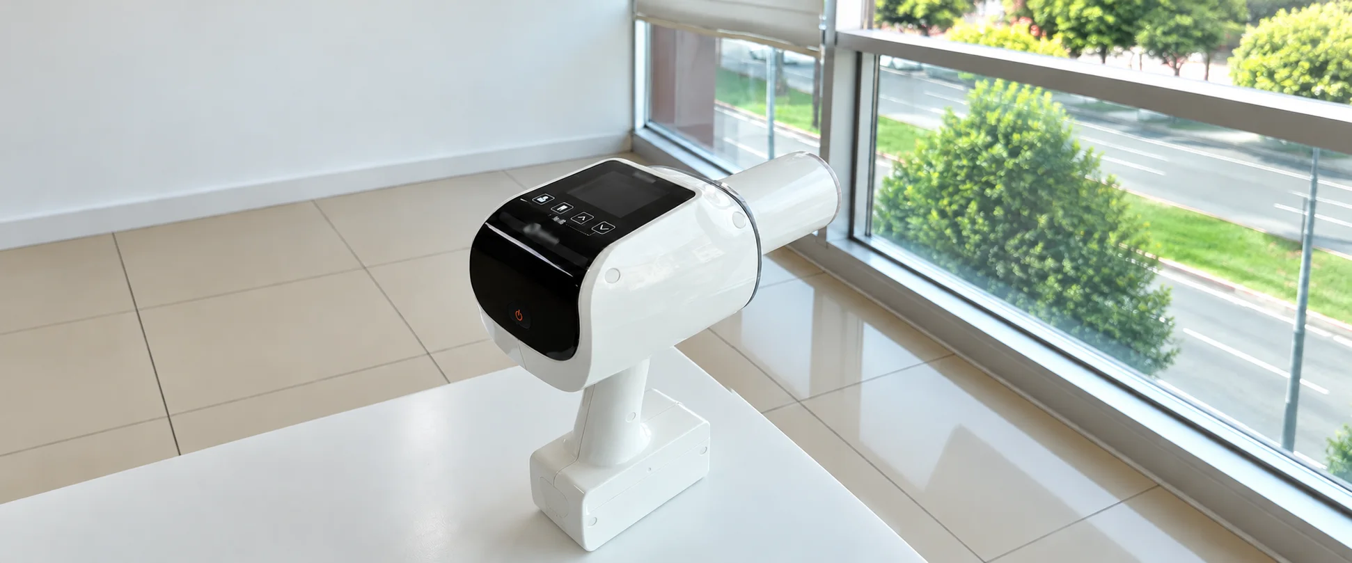
How are tooth roots and surrounding tissues imaged?

X-rays from the unit pass through the relevant area to a suitable sensor or receptor inside the mouth. The resulting two-dimensional image is assessed alongside clinical findings. Imaging is not needed at every check-up; the dentist decides according to clinical need.

  • Assessment of tooth roots
  • Examination of surrounding tissues
  • Imaging according to clinical need

Intraoral X-ray Unit

Intraoral X-ray Unit
Autogenous Tooth Graft Unit

How is tooth-derived graft material prepared?

A suitable tooth already scheduled for extraction may be processed into dentine-derived graft material through defined preparation steps. Sound teeth are not extracted for this purpose. The dentist assesses suitability based on the tooth, medical history and planned surgery.

  • Assessing tooth suitability
  • Preparation and processing steps
  • Individual surgical assessment

Autogenous Tooth Graft Unit

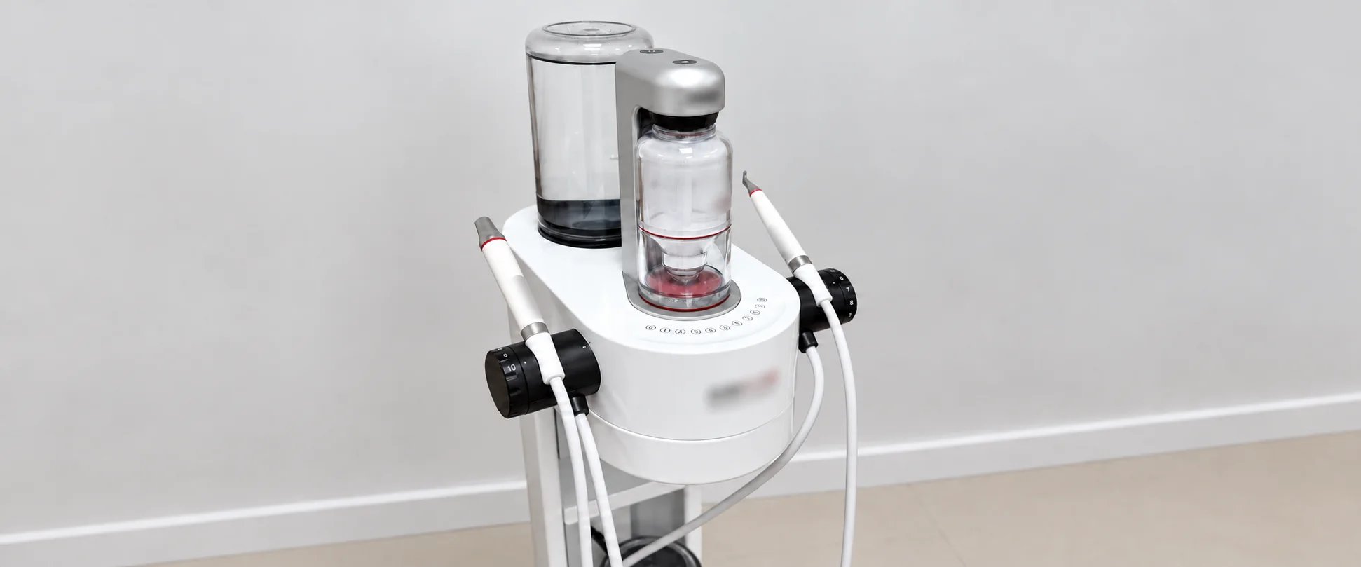
How are deposits on tooth surfaces removed?

Biofilm is a layer of microorganisms on teeth. After assessment, air, water and suitable powder cleaning or ultrasonic scaling may be used where needed. Methods and tips are selected for the teeth, gums and restorations; daily brushing and interdental care must continue.

  • Biofilm removal
  • External surface stains
  • Ultrasonic calculus removal

Biofilm and Calculus Removal Unit

Biofilm and Calculus Removal Unit

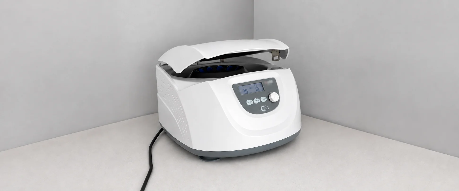
How are sample components separated?

The centrifuge spins tubes under controlled conditions, separating sample components by density. Preparation follows conditions suited to the sample and protocol. Having the device does not mean that a procedure is suitable for every patient or that it will produce a particular outcome.

  • Controlled rotational speed
  • Protocol-specific processing time
  • Separation of sample components

Centrifuge

Centrifuge

By section

Digital Workflow
1
Imaging
2
Surgery
1
General
1
Laboratory
1

All devices

Devices explained in detail appear above.

Digital Workflow

  • 01Intraoral Digital ScannerCreates a three-dimensional digital model by optically scanning teeth and surrounding oral surfaces. In suitable cases, it can be used to record impressions and share digital data with the laboratory. It is not an X-ray device and does not show the internal structure of roots or jawbone.see details

Imaging

  • 01Cone Beam Computed TomographyCBCTCreates three-dimensional cross-sectional images of the jaws and teeth. It may be used to assess bone structure, impacted teeth and nearby anatomical structures. It uses X-rays; the dentist determines whether a scan is needed and selects the imaging area.see details
  • 02Intraoral X-ray UnitProduces X-rays to image a specific area of the teeth, roots and surrounding tissues. The image is recorded with a suitable sensor or receptor placed inside the mouth. The dentist determines whether imaging is needed based on examination and existing images.see details

Surgery

  • 01Autogenous Tooth Graft UnitPrepares suitable teeth already scheduled for extraction into dentine-derived graft material for use in the same patient. It is not suitable for every tooth or surgical procedure; use depends on clinical assessment and the device’s operating conditions.see details

General

  • 01Biofilm and Calculus Removal UnitUses air, water and suitable cleaning powder to remove biofilm and external surface stains, and ultrasonic tips to remove calculus. The treatment area and method are selected after oral assessment. Professional cleaning does not replace daily oral care.see details

Laboratory

  • 01CentrifugeSeparates sample components by spinning tubes at a controlled speed for a set time. In clinical use, tubes, speed and duration are selected for the sample type and protocol. It is preparation equipment; its use alone is neither a treatment nor an indication of a treatment outcome.see details

Which device answers which question

How are digital impressions of oral surfaces taken?
Intraoral Digital Scanner
When is three-dimensional imaging of the jaws and teeth needed?
Cone Beam Computed Tomography
How are tooth roots and surrounding tissues imaged?
Intraoral X-ray Unit
How is tooth-derived graft material prepared?
Autogenous Tooth Graft Unit
How are deposits on tooth surfaces removed?
Biofilm and Calculus Removal Unit
How are sample components separated?
Centrifuge
ShareShare on WhatsApp

Let us call you back

Leave your details and our team will get back to you shortly.

Your details are never shared with third parties. We respond within 24 hours.

Cookies We use cookies to measure how the site is used. If you decline, the site works exactly the same; we simply do not measure.